OpenEPI · Tools
Structures, in the browser.
Type an RCSB PDB ID or open your own PDB or mmCIF file, and turn the structure in 3D: cartoon, surface and sticks, colored by chain, secondary structure or B-factor, with distances, hover labels and a PNG export.
Last updated September 17, 2026
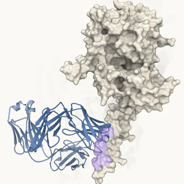
This viewer renders a macromolecular structure in the browser with 3Dmol.js, a WebGL library that runs entirely on your device: an entry is fetched from the RCSB Protein Data Bank by its four-character ID, or a local file is parsed in your browser and never uploaded anywhere. It is for anyone who wants to look at a target, an antibody or a peptide–receptor complex without installing software. Ligands, the HETATM records other than water, are drawn as sticks automatically.
Loading viewer…
Click two atoms to read the distance between their centers.
- Title
- —
- Chains
- —
- Ligands
- —
- Atoms
- —
- Source
- —
For reference and education. Nothing on this page is clinical guidance, a dose recommendation or an Epitorix performance figure; check every number against your own validated procedures.
How to read it
3Dmol.js 2.5
Representations and colors
- Cartoon
- The backbone trace: helices as ribbons, strands as arrows, the rest as tube. The default view.
- Surface
- The van der Waals surface at 60 % opacity over the cartoon, for the shape a ligand or a binder sees.
- Sticks
- Every heavy atom as a bond stick, for a close look at a site. Ligands are drawn as sticks in every mode, in violet under the house scheme.
- House
- Ink-gray protein, violet ligands: the site's palette, and the default.
- Chain
- One hue per chain, in the order the file lists them.
- Secondary structure
- Helix violet, sheet blue, coil gray, from the file's HELIX and SHEET records or 3Dmol.js's assignment when they are absent.
- B-factor
- Blue (lowest) to violet (highest) per atom, scaled within the loaded model. The B-factor is the crystallographic temperature factor; in cryo-EM entries the column holds whatever the depositors placed there.
Questions
files.rcsb.org
What the viewer does
- Where does the structure come from?
From the RCSB Protein Data Bank, the US site of the worldwide PDB archive, fetched at view time as the entry's PDB-format file, or its mmCIF file when no PDB-format file exists. The title, chains and ligand names in the summary are read from the entry's own header. The coordinates are not altered.
- Is my file uploaded?
No. A file you open is read into your browser's memory, parsed there and drawn there; nothing leaves the device, and closing the page discards it. The only network request the viewer makes is the download of a PDB entry you ask for, and that request carries only the four-character ID.
- What do the colors mean?
House: ink-gray protein with violet ligands. Chain: one hue per chain. Secondary structure: helix violet, sheet blue, coil gray. B-factor: a blue-to-violet gradient over each atom's temperature factor, scaled between the lowest and highest value in the loaded model, so the same color means different values in different entries.
- How does the distance measurement work?
In measure mode, click two atoms: the viewer draws a line between their centers and labels its length in ångström, from the deposited coordinates. For scale, a carbon–carbon single bond is about 1.5 Å, a hydrogen bond between two heavy atoms 2.7 to 3.2 Å, and a salt bridge is usually counted within 4 Å.
Sources and software
- Rego N, Koes D. 3Dmol.js: molecular visualization with WebGL. Bioinformatics 2015;31:1322–1324 (opens in a new tab) — the viewer library (BSD-3-Clause)
- 3Dmol.js — documentation and source (opens in a new tab) — representations, color schemes, selections
- Berman HM et al. The Protein Data Bank. Nucleic Acids Res 2000;28:235–242 (opens in a new tab) — the archive
- Burley SK et al. RCSB Protein Data Bank (RCSB.org): delivery of experimentally-determined PDB structures alongside one million computed structure models. Nucleic Acids Res 2023;51:D488–D508 (opens in a new tab) — RCSB PDB services and file downloads
- wwPDB consortium. Protein Data Bank: the single global archive for 3D macromolecular structure data. Nucleic Acids Res 2019;47:D520–D528 (opens in a new tab) — the archive's governance and CC0 data policy
- RCSB PDB — file download service (files.rcsb.org) (opens in a new tab) — the download URLs the viewer uses
- Peptide analyzer
- Kd to pKd and ΔG calculator
- From target to preclinical candidate
- Structures credited on this site
- Fab in the glossary
- All OpenEPI tools
A viewer, not an analysis. It draws deposited coordinates as they are; it does not predict, dock, score or validate anything, and nothing here is an Epitorix performance figure. Structure data: RCSB PDB (CC0 1.0); software: 3Dmol.js (BSD-3-Clause).